postmenopausal osteoporosis
Postmenopausal osteoporosis is a condition characterized by a decrease in bone density and an increased risk of bone fractures in women after they have gone through menopause. Menopause is a
natural biological process that marks the end of a woman's menstrual cycles, typically occurring between the ages of 45 and 55. During and after menopause, the levels of estrogen, a hormone
important for maintaining bone density, decline significantly. This can lead to a higher rate of bone loss and, consequently, postmenopausal osteoporosis.
Causes
-
Decline in estrogen levels: Estrogen helps to regulate bone remodeling, which is the process by which old bone tissue is replaced by new bone tissue. When estrogen levels drop
during menopause, the rate of bone resorption (the process of breaking down bone) increases, while the rate of bone formation decreases, leading to a loss of bone density.
- Age: As women age, they naturally lose bone mass. Postmenopausal osteoporosis is more common in older women because bone loss accelerates after menopause.
- Genetics: Some women may have a genetic predisposition to osteoporosis, making them more susceptible to bone loss.
- Lifestyle factors: Poor nutrition, lack of exercise, smoking, and excessive alcohol consumption can contribute to bone loss and increase the risk of osteoporosis.
Symptoms
Postmenopausal osteoporosis is often called a "silent disease" because it typically doesn't cause noticeable symptoms until a fracture occurs. Symptoms of fractures can include pain, swelling, and
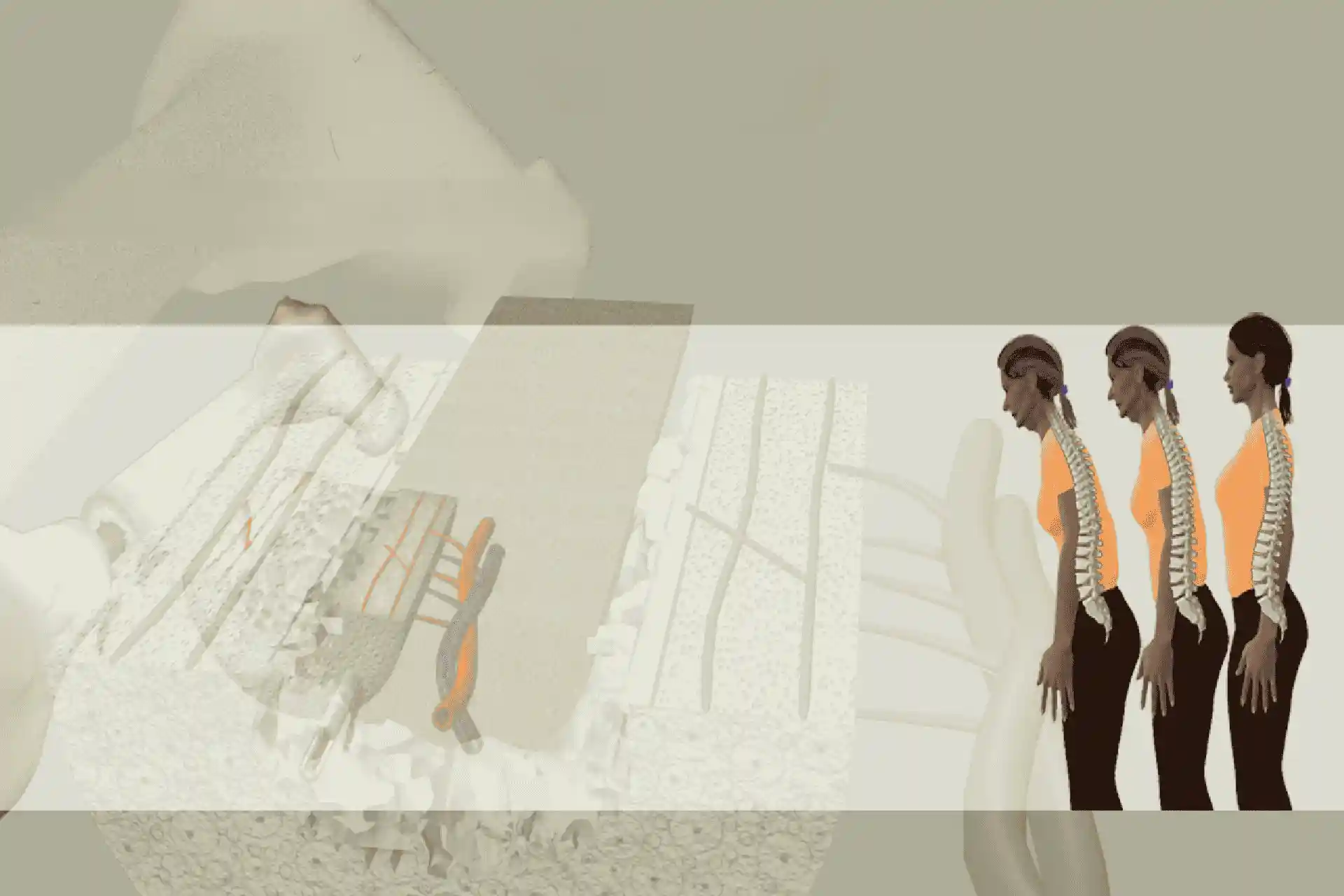
deformity at the fracture site. Common fracture locations include the spine, hip, and wrist.
Diagnosis
A bone mineral density (BMD) test, such as a dual-energy X-ray absorptiometry (DEXA) scan, is used to diagnose osteoporosis. The test measures the amount of bone mineral present in specific areas
of the body, and the results are compared to a reference population to determine if the bone density is within the normal range, low, or indicative of osteoporosis.
Treatment
Treatment for postmenopausal osteoporosis aims to slow bone loss, increase bone density, and prevent fractures. Options include:
-
Medications: Bisphosphonates, such as alendronate and risedronate, are commonly prescribed to help slow bone loss. Other medications, like denosumab, teriparatide, and
abaloparatide, can also be used depending on the severity of the condition and other factors.
- Hormone therapy: Estrogen replacement therapy, although not commonly recommended due to potential side effects, can help maintain bone density in some women.
-
Lifestyle changes: Maintaining a healthy diet rich in calcium and vitamin D, engaging in weight-bearing and resistance exercises, and avoiding smoking and excessive alcohol
consumption can help improve bone health.
-
Fall prevention: Reducing the risk of falls by making the home environment safer and improving muscle strength and balance can help prevent fractures associated with
osteoporosis.
It's important to note that early detection and intervention are crucial for managing postmenopausal osteoporosis and minimizing the risk of fractures. Regular check-ups and bone density tests can
help identify the condition early and allow for more effective treatment.
Transcript:-
Osteoporosis is a condition in which the density of the bones in the skeleton has reduced, making them weaker and prone to fracture globally. An estimated 9 million osteoporotic fractures occur each
year as life expectancy increases. So too will the annual number of osteoporotic fractures. In fact, a fourth pole rise is predicted over the next five decades.
Osteoporotic fractures cause pain, suffering, disability, and sometimes deformity. Osteoporosis is most commonly seen in women, although men can also be affect. For instance, in a old man in the uk, a
lifetime risk of osteoporotic hip fracture is 3.1%. In a woman, the risk is 11.4%. This presentation will focus on postmenopausal osteoporosis. Bone provides support for movement and protection of the
body's organs.
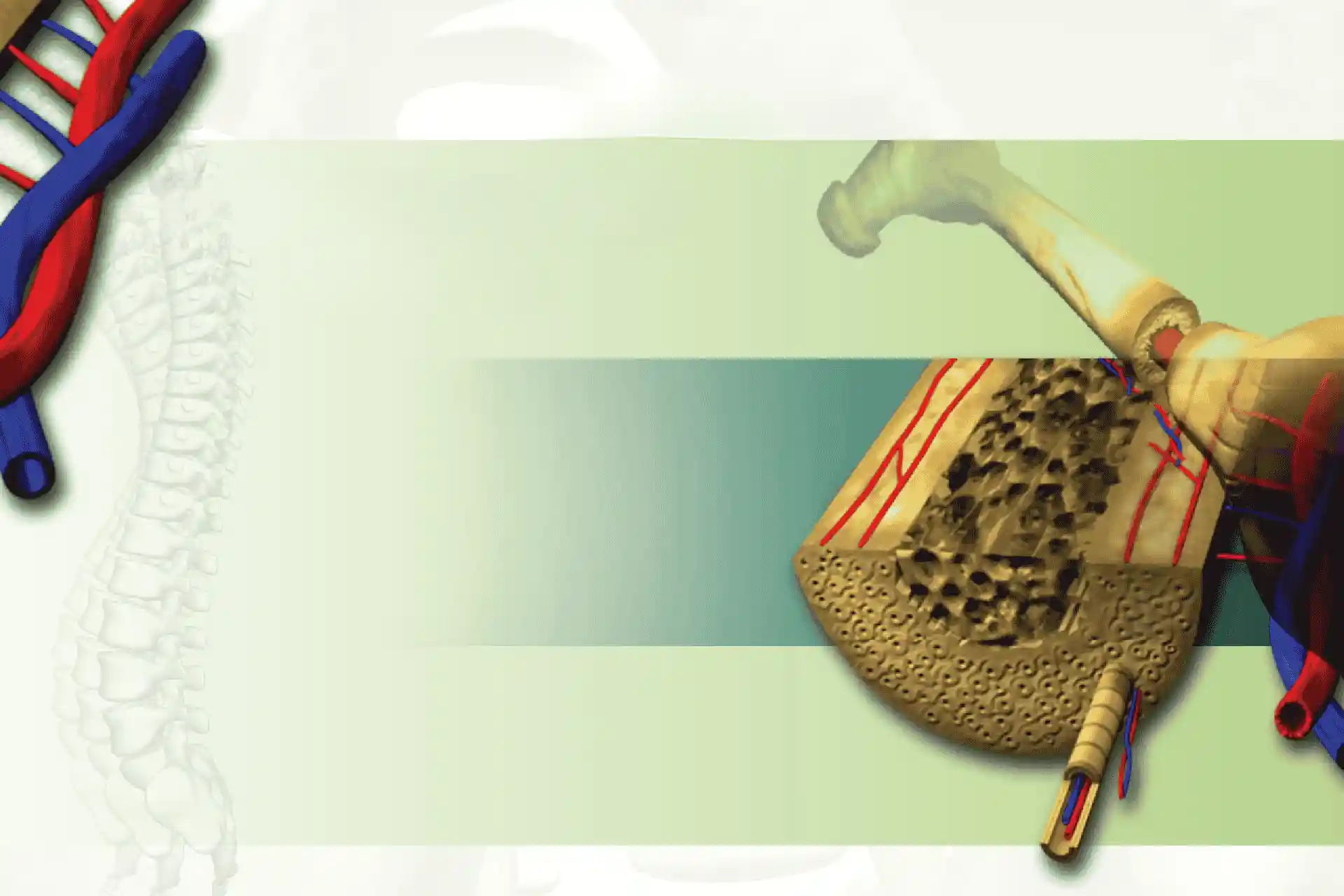
Ax as a reservoir of minerals such as calcium and produces blood cells. It has two components. The outermost cortical bone is dense and compact. Below. This is a spongy lattice, like trabecular bone
in the center, soft bone marrow, which produces blood cells, fills the holes and passageways of the. The combination of the dense combat, cortical and supple trabecular components make bones both
strong and light in weight. Bone is a living tissue. It is constantly in a state of renewal or repair Throughout our life, old bone tissue is replaced by new bone in a process called remodel. In the
first stage of remodeling called resorption, specialized bone cells, known as osteoclast, break down the bone.
Stage two sees new bone being formed by osteoblasts. The whole remodeling cycle takes about three to six months to complete throughout childhood, adolescence and young adulthood. The prime years of
physical growth, bone mass increases as bone formation exceeds resorption. Bone mass reaches its peak in the third decade.
Once peak bone mass is achieved, the balance shifts to a state of equilibrium where formation equals breakdown. Following menopause resorption speeds up. Since the female sex hormone, estrogen is no
longer present to keep resorption rate in. The underlying problem in post-menopausal osteoporosis, therefore is an imbalance in the remodeling process.
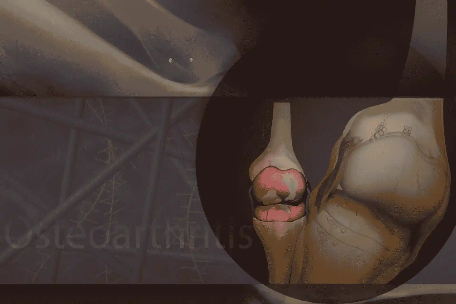
The negative balance in favor of resorption causes the bone mass to reduce, particularly in trabecular tissue. The spaces between the bone services become wider and the strength of the bone decreases.
The impact of this loss depends on how much bone is accumulated in the skeleton in childhood and early adult life. The peak bone mass osteoporosis is particularly common in women because they have a
lower peak bone mass than men. Hormonal changes after the menopause also accelerate the process of bone. Menopause or hysterectomy at an early age, therefore increases the risk of osteoporosis.
Advancing age and a family history of osteoporosis also increases the likelihood of the condition.
Finally, because of racial differences in bone density, osteoporosis is most common among Caucasians and Asian populations. Lack of deficiency of calcium may also be implicated as this mineral is
important for healthy bone. Vitamin D deficiency also increases risk as it is needed for the adequate absorption of calcium and mineralization of new bone.
Vitamin D deficiency is often seen in housebound individuals who are rarely exposed to sunlight. Excessive weight loss and dieting, lack of physical activity, smoking and alcohol. Overuse are other
factors that reduce bone density.
Finally, certain medications or medical conditions could cause bone loss and therefore predispose to the development of osteoporosis. These include long-term oral steroid therapy. Hyperparathyroidism,
intestinal mal-absorption, and rheumatoid arthritis.
In the early stages, there are usually no signs or symptoms of osteoporosis. This is why the condition is often described as silent. A fracture occurring after only mild trauma may be the first sign
that something is wrong. The most common fracture sites are the hips and wrist usually occurring due to minor falls and the vertebrae resulting from simple activities such as coughing, bending or
lifting.
Individuals with vertebra fractures may complain of back pain, loss of height. Or develop a stooped posture. Although these symptoms may occur with degenerative arthritis and disc disease, your doctor
will take a detailed medical history, perform a thorough physical examination, and take measurements of bone density before arriving at a conclusive diagnosis of osteoporosis.
Dual energy X-ray absorb geometry or dx A is typically used for bone density assessments at the hip or spine. A similar method measures bone density at the forearm, heel or hand. This is called
peripheral DX a or P D X A. Other machines that may be used to investigate your bone mass include quantitative computerized tomography or QC t. Quantitative ultrasound and radiographic absorption
treatment cannot cure osteoporosis, but instead aims to reduce the risk of fractures since these cause considerable pain, disability, and loss of independence.
Bisphosphonates are the most frequently used group of drugs for preventing and treating osteoporosis. They reduce the rate and extent of resorption leaving a positive balance during the remodeling
process. This increases bone mass over time and reduces the risk of fracture. Examples of bisphosphonate drugs are Aron. Nate, Ibandronate and Renate. If bisphosphonates are unsuitable, other types of
drugs exist. Other anti resorption drugs to prevent and treat osteoporosis include reen, calcitonin, and certain forms of hormone replacement therapy or H R T to replace the missing estro.
The health risks of long-term H R T may outweigh the benefits. So these drugs are no longer routinely used to help prevent osteoporosis in women aged over 50 years. However, H R T may be useful for
prevention in women with an early menopause where it may be continued until the average age of menopause, which is about 50 years. Parathyroid hormone or teraparatide affects bone resorption to a
small extent and increases bone formation. Promoting bone growth. Strum ranelate reduces resorption and stimulates bone formation. Both teraparatide and strontium ranelate are used to treat but not
prevent osteo por. Calcium and vitamin D supplements are often prescribed alongside these other forms of treatment.
Building strong bones early in life and maintaining a healthy lifestyle are the best defensed against osteoporosis, whatever your age or past history. There are several ways that you can lower the
risk of osteoporosis or preserve as much bone as possible during periods where mass is naturally lost over. Make sure that your diet contains enough calcium and vitamin D. Ask your doctor what levels
are recommended for your particular situation and where the supplements are advised.
Exposure to sunlight promotes the production of vitamin D in the skin. Get outdoors in the summer months to increase your vitamin D levels. 15 to 20 minutes twice a week during summer months is
recommended. Exercise regularly using weight-bearing activities such as brisk walking, jogging, or dancing, where your legs and feet support your body weight.
Avoid smoking and limit alcoholic drinks to a maximum of two per day. Try to lessen the chance of falls, for example, by getting your site checked and keeping the floor of your house clear from trip
hazard. Consult your doctor about falls if you have poor balance or a past history of falls. Ask your doctor for details of osteoporosis patient organizations as they can provide you with further
guidance and support. Finally, take your osteoporosis treatments if these are prescribed for you, as they can substantially reduce your risk of fracture.