LASIK and Cataract
LASIK
LASIK stands for Laser-Assisted In Situ Keratomileusis. It is a popular laser eye surgery procedure intended to correct vision problems like nearsightedness, farsightedness, and astigmatism. In
LASIK, a laser is used to reshape the cornea, the clear front cover of the eye, to improve visual focus.
How LASIK works:
- First, the eye surgeon uses a computer-controlled laser to cut a small flap in the cornea. The flap is then peeled back to expose the underlying corneal tissue.
-
The laser is then used to ablate or reshape the corneal tissue under the flap to correct vision deficiencies. Reshaping of the cornea changes the way light enters the eye, allowing for
improved visual focus.
- The corneal flap is then placed back to cover the ablated tissue. The flap adheres naturally without stitches.
- The procedure typically takes just a few minutes to complete, though the preparation and recovery time add several hours to the entire process.
- Vision recovery after LASIK can take several days to several weeks. Most patients return to normal activities within a few days.
LASIK (Laser-Assisted In Situ Keratomileusis)
LASIK is a type of refractive surgery that corrects common vision problems such as myopia (nearsightedness), hyperopia (farsightedness), and astigmatism. The goal of LASIK is to reduce or
eliminate the need for glasses or contact lenses.
The procedure involves using a specialized laser to reshape the cornea, which is the transparent front part of the eye. This reshaping helps to properly focus light onto the retina, resulting in
clearer vision. The LASIK procedure generally consists of the following steps:
- A protective corneal flap is created using a microkeratome (a precise surgical blade) or a femtosecond laser.
- The corneal flap is carefully lifted to expose the underlying corneal tissue.
- An excimer laser is used to remove a precise amount of corneal tissue, reshaping the cornea to correct the refractive error.
- The corneal flap is repositioned and adheres to the cornea without the need for stitches.
The procedure is fast, and recovery time is typically minimal, with most patients experiencing improved vision within 24 hours.
Cataract
A cataract is a clouding of the eye's lens, which is normally clear. Cataracts prevent light from properly focusing on the retina, resulting in blurred vision. Cataract surgery is a procedure to
remove the clouded lens and replace it with an artificial lens to restore vision.
How cataract surgery works:
- The eye surgeon makes a small incision in the cornea or white part of the eye.
- An ultrasound probe is inserted to break up and remove the clouded lens. This process is called phacoemulsification.
- An intraocular lens (IOL) implant is then placed in the empty lens capsule. The IOL provides the light focusing power of the natural lens.
- The incision is closed, and the procedure is complete. Like LASIK, vision may take some days or weeks to stabilize.
- Over 90% of people who have cataract surgery regain very good vision. The procedure is one of the safest and most effective surgeries performed today.
Cataract Surgery
Cataract surgery is performed to remove a clouded lens from the eye and, in most cases, replace it with an artificial intraocular lens (IOL). A cataract is a common age-related condition that
causes the natural lens of the eye to become cloudy, resulting in blurry or distorted vision.
Cataract surgery is one of the most common and effective surgical procedures performed worldwide. The procedure typically consists of the following steps:
- A small incision is made in the front part of the eye, near the edge of the cornea.
-
A technique called phacoemulsification is used to break up the cataract. This involves inserting a small ultrasound probe into the eye, which emits high-frequency sound waves to break the
cloudy lens into tiny fragments.
- The lens fragments are suctioned out through the probe.
-
An artificial intraocular lens (IOL) is inserted into the empty lens capsule to replace the natural lens. The IOL is usually made of plastic, silicone, or acrylic materials and is designed to
remain in place permanently.
- The incision is closed, and in most cases, no stitches are required.
Recovery from cataract surgery is generally quick, with most patients experiencing improved vision within a few days.
In summary, LASIK is a procedure that reshapes the cornea to correct refractive errors, while cataract surgery involves removing a clouded lens and replacing it with an artificial lens. Both
procedures are effective at improving vision, but they address different eye conditions.
Topic Highlights:-
- This visual presentation covers two topics, both LASIK & Cataract.
-
The first section on LASIK discusses in detail the anatomy of the eye, the function of the eye, refractory errors of the eye, correction with LASIK, advantages of LASIK, and steps to be taken
during recovery.
- The second section on Cataract deals with the condition in which the eye lens is clouded and affects vision, diagnosis of cataract, surgery, and recovery after surgery.
Transcript:-
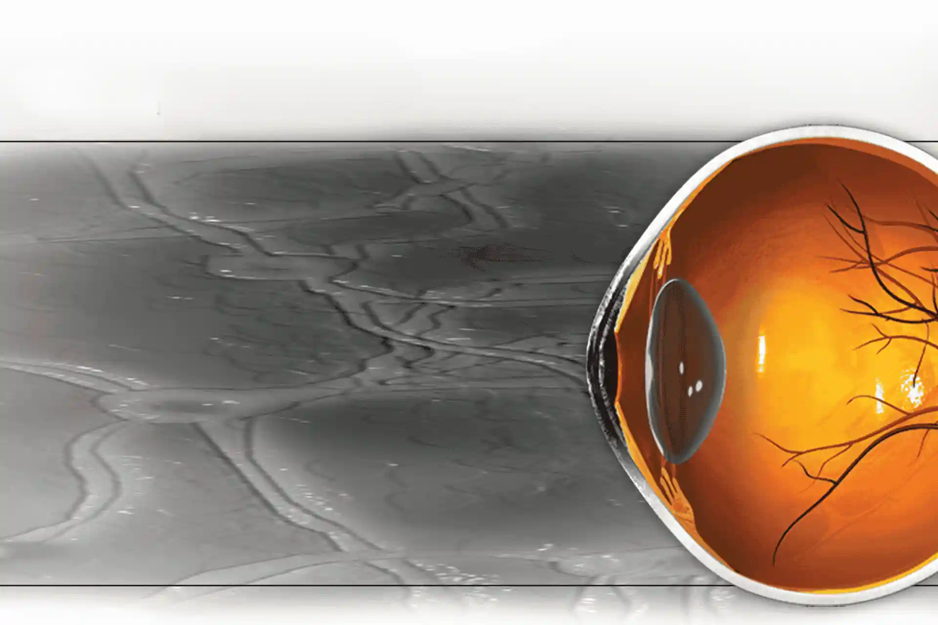
The eye is a very complex sense organ which responds to changes in light and enables us to see. The ability to see depends on various structures in the eye. The outermost part of the eye is the
eyelid, which protects the eye and lubricates the eyeball. The outermost layer of the eye is the sclera, the white of the eye. Muscles connected to the sclera control eye movements. The cornea is
transparent and covers the front of the eye. It is a powerful refracting surface where light rays are bent, refracted and focused.
The colored part of the eye is called the iris. It controls light levels inside the eye. The pupil is the round opening in the center of the iris. Dilator and sphincter muscles of the iris change the
pupil size according to the amount of light. The soft lens located behind the pupil allows the muscles to adjust its shape and focus. This allows us to focus on something close to us and far away. The
retina is a sensory tissue lining the back of the eye. The lens focuses light in the retina with the help of muscles attached to it. Photoreceptors in the retina capture light rays and convert them
into electrical impulses. The optic nerve carries these impulses to the brain.
Myopia, or short sight, is the inability to see distant objects as clearly as near objects. With myopia, the cornea is too steep, causing images to focus in front of the retina instead of directly on
it. Persons with hyperopia, or long sight, have more difficulty seeing near objects as clearly as distant objects. Hyperopia occurs when the cornea is too flat, causing images to focus behind the
retina. People with astigmatism have distortion of the image on the retina caused by irregularities in the cornea or lens of the eye.
There are many different treatments available depending on the individual’s needs. Glasses or contact lenses are designed to compensate for the eye’s imperfections. Several new techniques such as
LASIK offer millions of people the opportunity to reduce or eliminate their need for glasses or contact lenses. LASIK, or laser-assisted in situ keratomileusis, is a surgical procedure which can
correct a wide range of refractive errors such as myopia, hyperopia and astigmatism.
To be eligible for LASIK surgery, the patient should be over 18 and should not suffer from aliments such as diabetes and eye diseases. Patients should not be taking medications such as steroids. Women
should not be pregnant or breast-feeding. People with unstable vision are not eligible for LASIK. Vision should be stable at least for a year. People with large pupils are not eligible.
A baseline evaluation of the eye is done to decide whether the patient is a candidate for surgery. Current medical history is also discussed. When surgery is scheduled, patients are advised on
preparation for surgery. Contact lens wearers need to discontinue and switch to glasses at least 2 weeks prior to surgery in the case of soft contact lenses, 3 weeks in case of rigid gas permeable
lenses and 4 weeks in the case of hard lens. Cosmetics should be avoided the day before surgery.
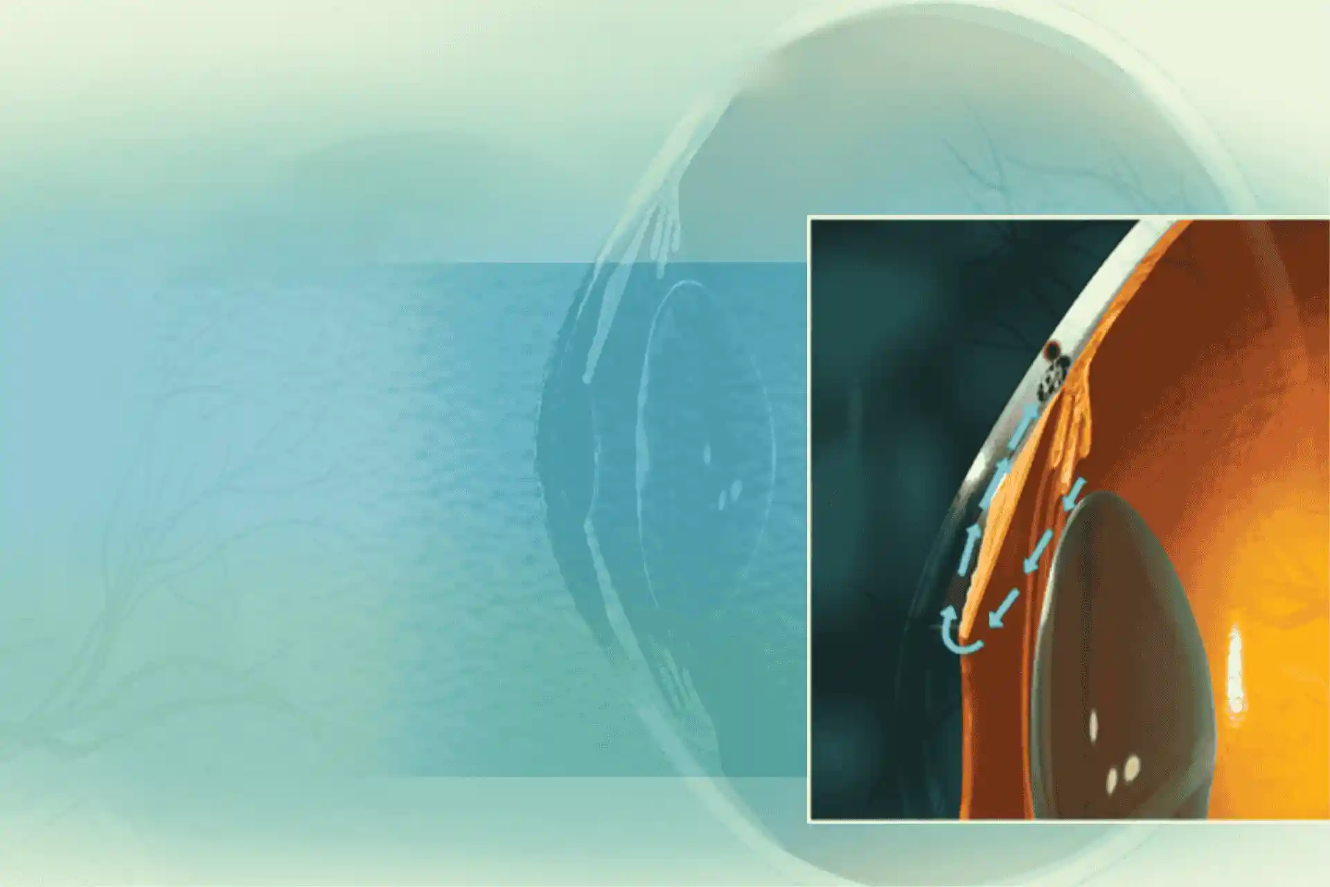
Before LASIK surgery, anesthetic drops are administered into the eyes. A suction ring is placed on the eye to stabilize and check the eye pressure. The microkeratome, a cutting instrument, is attached
to the suction ring. A thin flap of the cornea is lifted and the inner tissue of the cornea is exposed. The exposed layer of the cornea is then reshaped with an excimer laser. Each pulse removes a
microscopic layer. The actual laser treatment typically lasts less than one minute. The flap is folded back over the eye and the cornea heals naturally without the need for stitches.
To treat nearsightedness, the laser removes tissue from the center of the cornea, thereby making it flatter. In the case of farsightedness, the central part of the cornea is made steeper. This is done
by directing the laser beam to remove tissue from around this area. To treat astigmatism, the cornea is made more spherical. This is done by removing tissue in one direction more than the other by
changing the pattern of the beam.
Surgery usually takes less than 5 minutes for each eye. Patients often experience a blurring of vision immediately after the surgery. Patients are given eye shields to protect the eye from touching or
rubbing. Eye drops are administered to prevent eye infections. Vision improves and continues to clear. Pain is rare and mild, and goes away in a few hours. Patients may experience dry eyes,
sensitivity to light, and glare and halos, which normally subside as the eyes heal. Artificial tears are used to lubricate the eye. The patient should avoid contact sports and swimming for a couple of
days. The patient is also advised to avoid any lotions or creams near the eyes for two weeks following surgery. Strenuous work should be avoided the four weeks following surgery. Eyes are examined and
tested within the next two days.
While good vision is possible, expectations of perfection may be unrealistic. Some people require glasses for fine tuning vision for conditions such as night driving, computer work and heavy reading.
Complications of surgery, though rare, may occur in less than 1% of the patients. Overcorrection or undercorrections can happen occasionally. In this case the patient is prescribed contact lenses or
glasses. The flap could develop wrinkles, which may need repositioning. Some patients develop problems in night vision such as glare or halo. Permanent dry eye is another complication, which can be
treated with artificial tears. In rare cases repeat surgery may be done after 3 months by raising the same flap.
Cataract is clouding of the lens of the eye due to the deposition of proteins, which obstructs the passage of light. It can result in a partial or severe decrease in vision. Cataract is a significant
cause of low vision or blindness. Age related cataract is responsible for 48% of blindness in the world, which is nearly 18 million people. The number of people suffering from cataract is growing as
people live longer.
Cataract may cause cloudy, fuzzy or blurred vision and faded colors, halo around lights, double vision, and poor night vision. Cataracts usually progress gradually. Most age-related cataracts may
progress over a period of two years. Other types of cataracts, especially in younger people and diabetics, may progress faster.
The majority of cataracts are related to aging, although cataracts are sometimes seen in babies. This type of cataract is called congenital cataract. Cataracts may also develop as a result of other
diseases such as diabetes or trauma to the eye. Medications such as steroids and long-term exposure to sun may cause cataracts.
Cataracts are classified according to the location on the lens. Nuclear cataract is usually associated with aging and is the most common type of cataract. Nuclear cataracts develop in the center of
the lens. Cortical cataract develops in the lens cortex, and gradually spreads its wedge shaped spokes towards the center of the lens. This is more common among people with diabetes. It may impair
both distance and near vision and may require surgery at early stages. Subcapsular cataract develops at the back of the lens. People with diabetes, high myopia and those taking high doses of steroids
are more susceptible.
Your doctor will examine the eyes thoroughly. Glare testing and contrast sensitivity tests are sometimes done. Cataracts can only be removed with surgery and the decision for surgery depends on the
loss of vision. Even after surgery problems such as macular degeneration, glaucoma and diabetes may affect vision in some people. The chances of restoring good vision following the surgery are
excellent depending on the health of the eyes.
Anesthetic drops are used to numb the eye and a small incision about 3mm is made on the cornea. Special microsurgical instruments break the cataract in a process called phacoemulsification and suck
the fragments of the lens from the eye. The back portion of the lens capsule is left intact to support the small foldable intraocular lens, which is folded and inserted through the wound. It is then
opened and implanted in place of the natural lens. The lens is held in place by the arms of the lens, known as haptics. The incision is self-sealing so no stitches are needed.
Patients may be asked to wear a shield to protect the eye, especially while sleeping. Avoid rubbing the eyes during the first few weeks following surgery. Antibiotic and anti-inflammatory eye drops
will be administered during the first few days. Your surgeon may prescribe new glasses after vision has stabilized. This may take four to six weeks. Patients can resume normal activities following
surgery. Patients may read, watch television or do light work.
Cataract surgery is generally safe. In rare cases infection, inflammation or bleeding may occur in the eye following surgery. Dislocation of the implanted lens and retinal detachment are also possible
complications. A thickening of the membrane behind the lens known as after cataract may occur months later.